Basal cell carcinoma is the most common form of skin cancer. This tumor is not life threatening, except in the rarest of circumstances when tumors go untreated for decades. For most, basal cell carcinoma grows slowly over years, and appears as a pimple-like growth that bleeds, heals, and re-bleeds. If untreated, basal cell carcinoma my penetrate and destroy deeper tissues and can be mutilating.
Most basal cell carcinomas are caused by ultraviolet rays from the sun. It is known that chronic sun exposure, radiation, and tanning bed use can lead to the development of basal cell carcinomas.
Dr. Bader was invited to author the chapter regarding basal cell carcinoma on the online medical reference textbook on emedicine.com, a subsidiary of WebMD. To see this article please click here: Basal Cell Carcinoma.
There are different histologic subtypes of basal cell carcinoma. These types are classified based on growth patterns that can be indentified under the microscope. Some types are more difficult to treat and do not respond as well to all treatment options. Therefore, one must take into account several factors when choosing an appropriate treatment or even the option not to treat. Such factors include: the age of the patient, the location of the tumor (i.e. is it close to the eye), the overall health of the patient (i.e. are able to undergo surgery), medications (i.e. are they on blood thinners that they are unable to stop, the histologic subtype of the tumor, the ability and desire of the patient to care for the site after treatment, the importance of a good cosmetic outcome after treatment, and cost.
Histologic Subtypes of Basal Cell Carcinoma:
There are several different types of basal cell carcinoma that are categorized by how they look under the microscope. Here are the most common types:
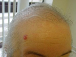
Nodular Basal Cell Carcinoma is the most common type of basal cell carcinoma. This tumor usually looks like a pimple that bleeds, heals, and then re-bleeds. With time, these tumors enlarge and usually ulcerate (form an open sore) usually in the center. Under the microscope, these tumors grow as large, ball-like collections (or nodules), hence the name nodular basal cell carcinoma. This type of tumor is often treated with excision, electrodesiccation & curettage, Mohs’ micrographic surgery, or radiation.
Superficial Basal Cell Carcinoma is common, and looks like a pink patch of skin that may have some scale and may have one or more tiny scabs. This type of basal cell is often misdiagnosed as eczema, ringworm, or psoriasis. The tumor cells are attached to the undersurface of the epidermis, the top layer of skin. Hence, it has been termed a superficial basal cell carcinoma. Treatment for this type of basal cell carcinoma is similar to those listed above for Nodular basal cell carcinoma, although topical creams, such as Aldara or 5-Fluorouracil, can be used.
Infiltrating Basal Cell Carcinoma grows as strands of tumor cells the grow (infiltrate) between the collagen fibers within the dermis. For this reason, it is more difficult to identify the margins of the tumor clinically (by looking). These tumors do not have the typical pimple-like appearance of a nodular basal cell. These tumors are best treated using Mohs’ Micrographic Surgery, although wide excision and radiation are often used.
Morpheaform or Sclerosing Basal Cell Carcinoma often appears as a waxy, yellowish area or a scar. As they do not have the typical appearance of a nodular basal cell, rarely ulcerate, and look rather harmless, they are often not detected by the patient and non-Dermatologists. Often, these tumors are much larger than they appear, making treatment more difficult than other types of basal cell carcinoma. For this reason, Mohs’ Micrographic Surgery is the treatment of choice.
Read about the treatment for Basal Cell Carcinoma on Medscape by clicking here.